Retinopathy of Prematurity
Type: patient education, full color
Objective: Illustration is part of a series of illustrations for a poster depicting Retinopathy of Prematurity. Physicians will utilize this poster while explaining the condition of ROP to parents of newborns.
© 2020 Melissa Wallin. All rights reserved.
Acrocyanosis in Newborns
Type: educational, full color
Objective: Illustration was created to accompany a question testing nursing students’ understanding of and ability to identify acrocyanosis in newborns.
© 2020 Melissa Wallin. All rights reserved.
“Rare Genetic Diseases: Nature’s Experiments on Human Development,” Lee, C. E., Singleton, K. S., Wallin, M., & Faundez, V. iScience, 2020, https://doi.org/10.1016/j.isci.2020.101123
Figure for Rare Genetic Diseases Article
Type: figure for journal publication, full color
Objective: This illustration was created for a client to supplement the text of a publication.
© 2020 Melissa Wallin. All rights reserved.
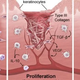
Stages of Wound Healing
Type: educational, full color
Objective: Illustration was created to accompany a USMLE question testing physicians’ understanding of the factors involved in the progression of wound healing.
© 2020 Melissa Wallin. All rights reserved.
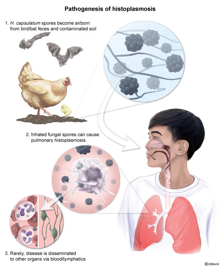
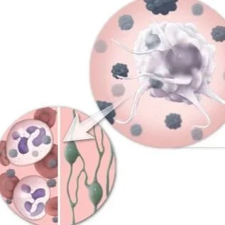
Pathogenesis of Histoplasmosis
Type: educational, full color
Objective: Illustration was created to accompany a question testing nursing students’ understanding of the development of histoplasmosis.
© 2019 Melissa Wallin. All rights reserved.
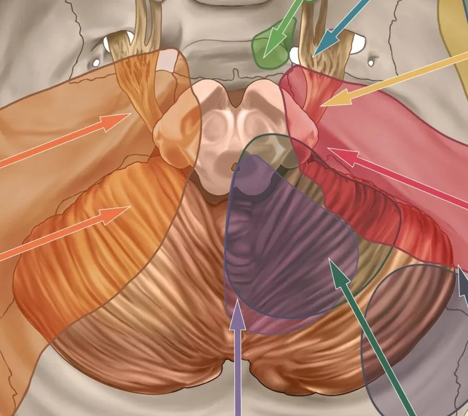
Images depict surgical approaches for brainstem access and accompany text in a chapter of the Neurosurgical Atlas.
Image 1 of 2 for Brainstem Corridors (Image 1)
Type: figure for surgical atlas, full color
Image 2 of 2 for Brainstem Corridors (Image 2)
Type: figure for surgical atlas, full color
© 2019 Melissa Wallin. All rights reserved.
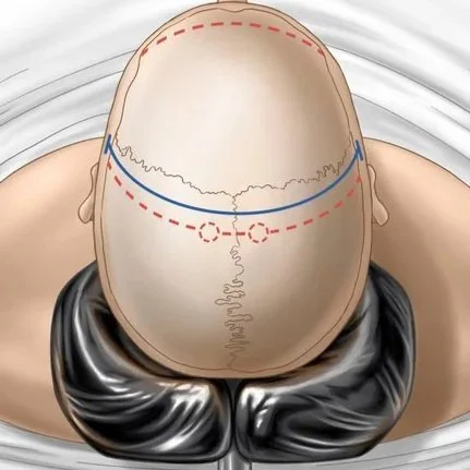
Images depict surgical technique used for a decompressive craniectomy and accompany text in a chapter of the Neurosurgical Atlas.
Image 1 of 3 for Decompressive Craniectomy (Image 1)
Type: figure for surgical atlas, full color
Image 2 of 3 for Decompressive Craniectomy (Image 2)
Type: figure for surgical atlas, full color
Image 3 of 3 for Decompressive Craniectomy (Image 3)
Type: figure for surgical atlas, full color
© 2019 Melissa Wallin. All rights reserved.
Objective: This illustration was created to enable medical students to better understand a challenging view of the internal structures of the basal ganglia and as a result correctly identify the structures in radiology and dissection lab.
Structures of the Basal Ganglia in the Axial Plane (Image 1)
Type: figure for anatomical atlas, pen and ink
Structures of the Basal Ganglia in the Axial Plane (Image 2)
Type: figure for neuroanatomy lab manual, full color
© 2019 Melissa Wallin. All rights reserved.
Objective: Present the salient details of the repair of the injury and post-operative condition of the plaintiff so that a specialist can utilize the board to explain those details to a jury of laypeople.
Board 1 of 2 for Medical Legal Plaintiff Case (Image 1)
Type: medical legal, full color
Board 2 of 2 for Medical Legal Plaintiff Case (Image 2)
Type: medical legal, full color
© 2019 Melissa Wallin. All rights reserved.
Plate 1 of 2 Depicting Mitral Valve Repair Surgery (Image 1)
Type: surgical, tonal
Objective: To depict the steps of a surgical procedure so that a surgical intern or resident is able to understand the basic steps and able to assist. Steps depicted in this plate include (A) a right atriotomy (B) a left atriotomy (C) mitral valve exposure.
Plate 2 of 2 Depicting Mitral Valve Repair Surgery (Image 2)
Type: surgical, tonal
Objective: To depict the steps of a surgical procedure so that a surgical intern or resident is able to understand the basic steps and able to assist. Steps depicted in this plate include (D) suturing the annulus (E) suturing the annuloplasty ring (F1) annuloplasty ring placement (F2) suture fastening.
© 2019 Melissa Wallin. All rights reserved.
Vasculature of Kidneys and Adrenal Glands
Type: anatomical textbook, full color
Objective: This illustration was created to aid medical students in learning the blood supply to and from the kidneys and adrenal glands. After reviewing this piece, students should be able to identify the vasculature structures on a cadaver during an exam.
© 2019 Melissa Wallin. All rights reserved.
Objective: To be utilized by physicians, clinicians, and counselors, the purpose is to educate patients 15-25 YOA about how ADHD (Attention Deficit Hyperactivity Disorder), its treatments and management, and related brain regions are involved. The role-playing game (RPG) style attracts young adults’ attention by utilizing elements seen in games to bring a sense of adventure and approachability to the intimidating topic of brain anatomy and disorder treatment.
Your Journey Through ADHD Wall Poster (Image 1)
Type: wall poster for patient education, full color
Front Folds of Patient Education Brochure (Image 2)
Type: patient education trifold brochure, full color
Back Folds of Patient Education Brochure (Image 3)
Type: patient education brochure, full color
© 2019 Melissa Wallin. All rights reserved.
Portrait study, Long pose figure drawing, Cast drawing study
Type: graphite and white charcoal on tonal paper
© 2019 Melissa Wallin. All rights reserved.